
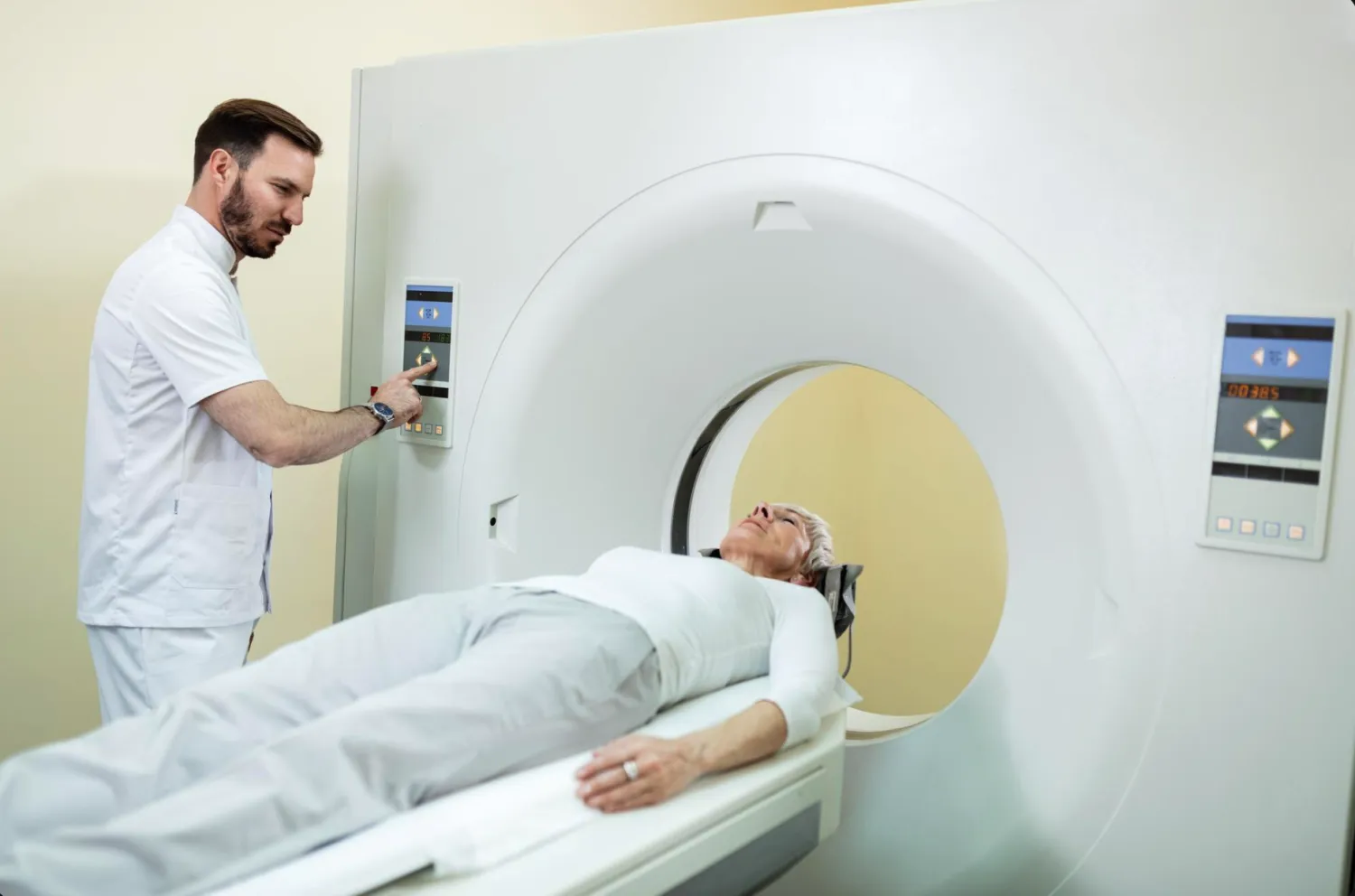
Both MRI and CT scans give detailed pictures of the inside of the body, but they work in different ways and suit different situations. Your doctor will choose the one that answers your question best.
How they work
- CT (computed tomography): uses a series of X-rays and a computer to build cross-section pictures. It is fast, usually taking only a few minutes.
- MRI (magnetic resonance imaging): uses a strong magnet and radio waves, with no radiation. It takes longer, often 20 to 45 minutes, and the machine is noisy.
When each is commonly used
- CT is often used for emergencies, injuries, bones, the lungs, chest and belly, because it is quick and clear.
- MRI is often used for the brain, spine, joints, ligaments and other soft tissues, where fine detail matters.
Safety and preparation
CT uses a small dose of radiation, so tell the team if you may be pregnant. MRI uses no radiation but the magnet can affect some metal implants, so tell the team about any pacemaker, implants or metal in your body. Some scans use a contrast dye, and you may be asked about allergies or kidney problems.
Which one do you need?
That depends on what your doctor wants to look at. If you are not sure, ask them why a particular scan was chosen, and follow the preparation steps for your appointment.
This article is general information and not medical advice. Please talk to your doctor about your own results and health.